- Home
- Shoulder Anatomy
- Shoulder Bones
Shoulder Bones
Written By: Chloe Wilson BSc (Hons) Physiotherapy
Reviewed By: SPE Medical Review Board
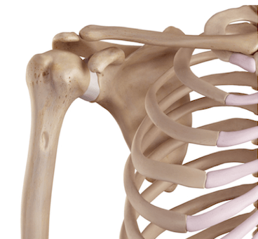
The shoulder bones fit together to form a highly complex, highly mobile joint that allows our arms to move through a huge range in multiple directions. In fact, the shoulder is the most mobile joint in the entire human body.
It is also the least stable joint so relies on a complex network of ligaments, tendons and muscles to function effectively.
Whilst shoulder bone injuries are much less common that soft tissue injuries, fractures, dislocations and shoulder bone spurs can cause significant problems.
Here we will look at the different aspects of shoulder bone anatomy including each of the four shoulder joints, the surrounding soft tissues and common problems that can affect the shoulder bones.
Shoulder Bone Anatomy
There are three shoulder bones:
- Humerus: aka upper arm bone
- Clavicle: aka collarbone
- Scapula: aka shoulder blade
There are a number of different muscles, tendons and ligaments that attach to the shoulder bones that work together to provide stability and strength to this highly mobile joint so we can lift and carry things, throw, push and pull and perform many other actions with our arms.
1. Upper Arm Bone
The first shoulder bone we are going to look at is the upper arm bone, also known as the humerus bone.
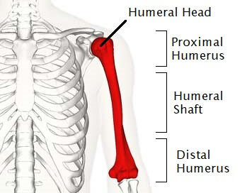
The humerus is a long bone that runs between the shoulder and the elbow. It is the bone found in the upper arm and is the longest bone in the upper limb.
At the top of the upper arm bone is the head of humerus, a rounded area that resembles a ball. The head of humerus rests in a shallow socket in the shoulder blade forming the shoulder joint.
The humerus can be divided into three sections:
- Proximal Humerus: the top section of the upper arm bone comprising of the head and neck of the humerus
- Humeral Shaft: the mid-section of the long humeral shoulder bone
- Distal Humerus: The lower section of the humerus, near the elbow
Key Function: the ball shaping of the head of humerus allows the shoulder to move through a huge range of motion, more than any other joint in the body.
2. Scapula Bone
The scapula, aka shoulder blade, is another important shoulder bone that forms the socket part of the shoulder joint.
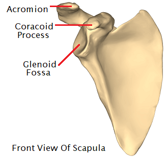
The shoulder blades are large, flat, triangular shaped bones found at the back of the shoulder, lying over the upper rib cage. The outer corner is shaped into a shallow, inward facing dome, known as the glenoid fossa, which forms a shallow socket for the ball shaped head of humerus.
There are three distinct areas on the scapula bones that stick out forming important bony prominences for the attachment of muscles, tendons and ligaments:
- Acromion Process: on the top, outer corner of the scapula bone
- Coracoid Process: sticks out at the front of the scapula bone
- Spine Of Scapula: a ridge that runs across the back of the scapula bone
Fun fact, no less than 18 different muscles attach to the scapula bone.
Key Functions: The scapula bone helps stabilise the upper arm bone and forms the socket for the shoulder joint
3. Clavicle Bone
The clavicle bone is the front-most shoulder bone, also known as the collar bone.
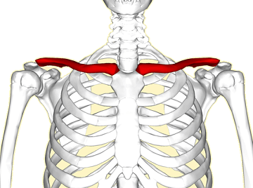
The clavicle is a long, thin bone that connects the shoulder to the chest via the sternum and is the only long bone in the human body that sits horizontally.
Located directly above the first rib, the clavicle shoulder bone acts like a crane to support the shoulder blade in place so that your arm can hang down freely by your side and swing as you walk.
Fun fact – the clavicle is the first bone to start developing in embryos but one of the last to full develop, often not reaching full ossification until people are in their early 20s.
Key Function: the clavicle forms the roof of the shoulder joint and transmits forces between the arm and the trunk
Shoulder Joint Anatomy
The shoulder is often thought of as being just one joint, but the three shoulder bones actually join together in different places to form four different shoulder joints:
- Glenohumeral Joint: The ball and socket articulation between the upper arm bone and shoulder blade. This is the joint classically referred to as “the shoulder joint”
- Acromioclavicular Joint: the joint between the collar bone and the shoulder blade via the acromion process
- Sternoclavicular Joint: the joint between the collar bone and the chest bone
- Scapulothoracic Joint: the large articulation at the back of the chest between the shoulder blade bones and the underlying rib cage.
You can find out more about each one in the shoulder joint anatomy section.
Structures Around The Shoulder Bones
The shoulder bones are connected to and surrounded by various structures that help the shoulder joint to function effectively including:
1. Shoulder Muscles
A number of muscles surround the shoulder bones and joints, working together to provide movement, support and stability to the shoulder. The two main groups of muscles that attach to the shoulder bones are the:
- Rotator Cuff: Four muscles that surround the shoulder joint - supraspinatus, infraspinatus, teres minor and subscapularis.
- Biceps & Triceps: the main muscles of the upper arm connecting the elbow and shoulder bones
Each of the muscles attach to the various shoulder bones via tendons - strong, flexible cord like structures. It is really important to have good strength in the shoulder muscles to ensure the shoulder has adequate support - rotator cuff strengthening and scapular stabilisation exercises can really help.
Common Problems: torn rotator cuff, biceps and triceps tendonitis, rotator cuff tendonitis
2. Shoulder Ligaments
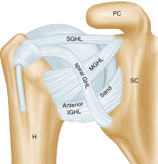
The shoulder ligaments and tough, elastic bands of connective tissues that connect bone to bone. The shoulder ligaments play a vital role in shoulder stability, supporting the joints and limiting joint movement.
There are nine different shoulder ligaments that connect the various shoulder bones at the glenohumeral, acromioclavicular and sternoclavicular joints.
Common Problems: Over-stretching, tearing or degeneration of the shoulder ligaments, usually due to an injury or repetitive friction
3. Shoulder Bursa
Bursa are small, fluid filled sacs that sit between the shoulder bones and tendons to reduce friction and allow smooth movement. There are a number of different bursa located around the shoulder, including the subacromial, subscapular and subdeltoid bursa.
Common Problems: Inflammation of the bursa known as shoulder bursitis
4. Shoulder Labrum
The labrum is a special layer of cartilage that lines the outer rim of the shoulder socket on the scapula bone. The glenoid labrum helps to deepen the socket which improves the congruity of the ball and socket joint, improving shoulder stability.
Common Problems: Damage to the labrum results in shoulder labrum tears which can lead to ongoing shoulder pain and instability
Common Shoulder Bone Problems
Damage to the shoulder bones from injuries or wear and tear are a common problem. The most common injuries affecting the shoulder bones are:
1. Shoulder Fractures
If enough force is placed through the shoulder, then one or more of the shoulder bones can break. Shoulder bone fractures are usually caused by:
- A Fall: typically either landing directly onto the shoulder or onto an outstretched arm
- Direct Blow: to the shoulder bones e.g. road traffic accident or sporting tackle
The most common fractures to affect the shoulder bones are:
- Proximal Humerus Fracture: a break in the top part of the upper arm bone affecting the ball shaped head of humerus. These are the most common shoulder bone fractures in adults
- Clavicle Fractures: a break anywhere along the length of the collar bone. The clavicle is the most frequently broken shoulder bone in children
Other possible fractures of the shoulder bones include:
- Scapular Fractures: extremely rare
- Humeral Shaft Fractures: in the mid-region of the upper arm bone, below the shoulder joint
2. Shoulder Impingement Syndrome
Primary shoulder impingement is a common problem caused by bony abnormalities at the shoulder.
Changes in the shape or angle of the acromion, typically due to bone spurs (small outgrowths of bone) reduce the space between the shoulder bones at the top of the glenohumeral joint. This increases the friction on the soft tissues leading to inflammation and pain, which can limit shoulder movement, and is known as shoulder impingement syndrome.
Common symptoms of shoulder impingement syndrome include:
- Painful Arc: the classic sign of shoulder impingement – increasing pain as you raise your arm above shoulder height that then decreases the higher you lift. Shoulder impingement pain tends to be worst between around 70-120 degrees of shoulder abduction
- Shoulder Pain: pain across the shoulder which may extend down the upper arm. Typically a sharp or toothache type pain. Particularly bad when reaching up or behind your back
- Daily Activities Affected: particularly sleeping and lifting heavy objects
If your doctor suspects impingement syndrome you may be sent for x-rays to identify any spurs growing on your shoulder bones that may need to be surgically removed with a subacromial decompression.
Related Article: Shoulder Impingement - Causes, Symptoms & Treatment
3. Shoulder Arthritis
Shoulder arthritis is caused by inflammation in one or more of the shoulder joints. The most common type of arthritis to cause shoulder pain is osteoarthritis.
Shoulder osteoarthritis causes gradual wear and tear of the shoulder bones, damaging the protective cartilage and causing it to wear away, fray and even tear. This reduces the space between the shoulder bones causing them to rub against each other which, in time, leads to pain and stiffness that can limit shoulder movement and daily activities.
Osteoarthritis is a particularly common problem affecting the shoulder bones in people over the age of 50. It can affect any of the shoulder joints, but is most common in the acromioclavicular joints.
Summary
The anatomy of the shoulder comprises of three shoulder bones, the humerus, clavicle and scapula, between them four different shoulder joints.
The shoulder bones are surrounded by muscles, tendons, ligaments and bursa. Having good strength and stability is vital to support highly mobile shoulder joint and it can really help to do:
Shoulder bone injuries are less common than soft tissue injuries. The most common shoulder bone problems are:
- Shoulder Fractures: broken bone
- Shoulder Impingement: from bone spurs
If you have pain from a problem in your shoulder bones or the surrounding soft tissues and want help finding out what is wrong, check out our shoulder pain diagnosis charts.
You may also be interested in the following articles:
Related Articles
Medical & Scientific References
- National Library Of Medicine: Anatomy, Shoulder and Upper Limb
- Medscape: Shoulder Joint Anatomy
- Arthritis Foundation: Shoulder Anatomy
- Radiopaedia: The Shoulder
- Journal Of Athletic Training: Functional Anatomy Of The Shoulder
Page Last Updated: December 10th, 2025
Next Review Due: December 10th, 2027
